Dr. Ritu Tiwari is an experienced Oral and Maxillofacial Radiologist with over ten years of expertise in diagnostic imaging in the fields of dentistry and healthcare. She completed her residency in this specialty at the University of California, Los Angeles (UCLA) and earned a master’s degree in Oral Medicine and Radiology from India. Dr. Tiwari has authored numerous peer-reviewed articles in oral medicine and radiology, with her work garnering over 100 citations. Her significant contributions to the discipline have been recognized through prestigious accolades, including the 2021 Albert G. Richards Research Award from the American Association of Oral and Maxillofacial Radiology (AAOMR). Presently, Dr. Tiwari serves as an Assistant Professor at the University of Texas Health Science Center at Houston, where she actively engages in both teaching and clinical radiology practice.
Credentials: BDS, MDS, Cert. Academic appointment: Assistant Professor, UT Health Houston Professional affiliations: AAOMR, AAOMFR, ADEA NPI: 1659094902
Detailed radiology reports enhanced with clinically relevant images.
Condensed CBCT reports
Image-free, diagnosis-driven reports designed to speed up clinical decisions
2D imaging
Radiology reports for dental imaging, including panoramic and full mouth series.
Comparison with prior scans
Comparison with prior studies to track progression and treatment response.
Second-opinion
TMJ, MRI, and MDCT second reads with focus on dental and occlusal considerations.
Trusted by Dental Professionals
Scans interpreted
0+
Turnaround time
48 - 0h
Case Studies
Explore some diagnostic radiology findings that were identified on routine CBCT scans. Each case emphasizes radiological observations that influence diagnosis, treatment planning and referral recommendations.
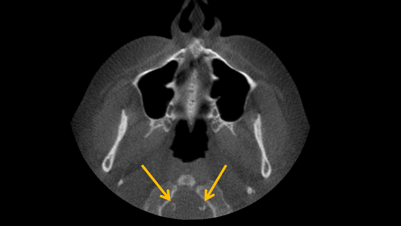
Study 1
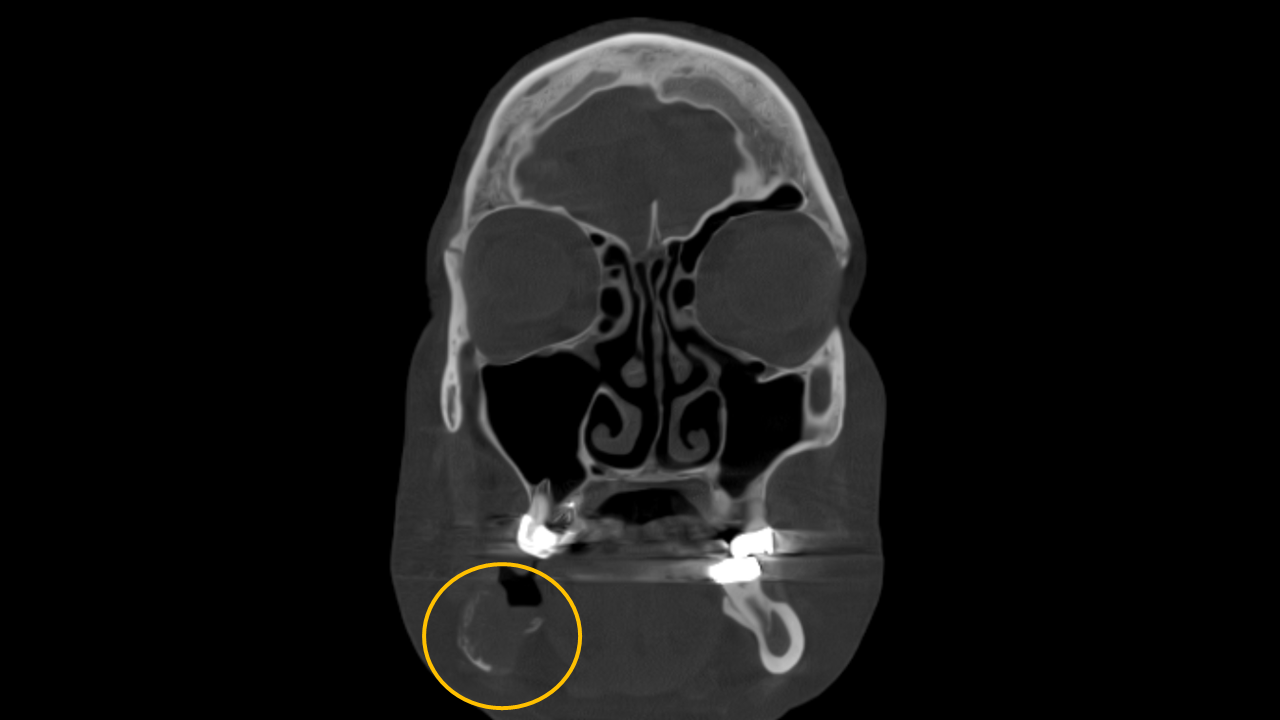
Finding: Circumferential radiodensities within the soft tissues posterior to anterior arch of atlas representing vertebral artery calcification.
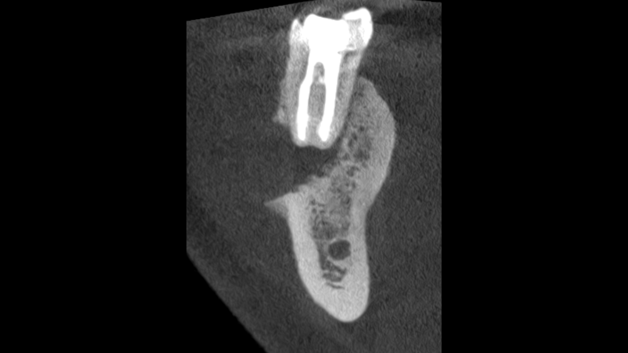
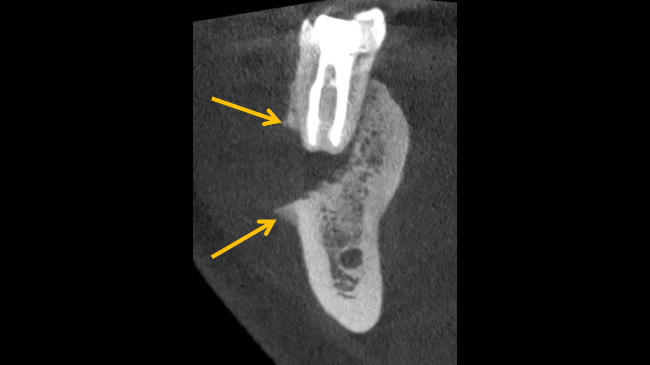
Study 2
Finding: A low-density, destructive bony lesion is associated with endodontically treated tooth #30 and has eroded the adjacent buccal cortex. Codman triangle is evident at the buccal cortex, and the surrounding trabeculation appears sclerotic. These imaging findings are suggestive of a chronic inflammatory process or malignancy.
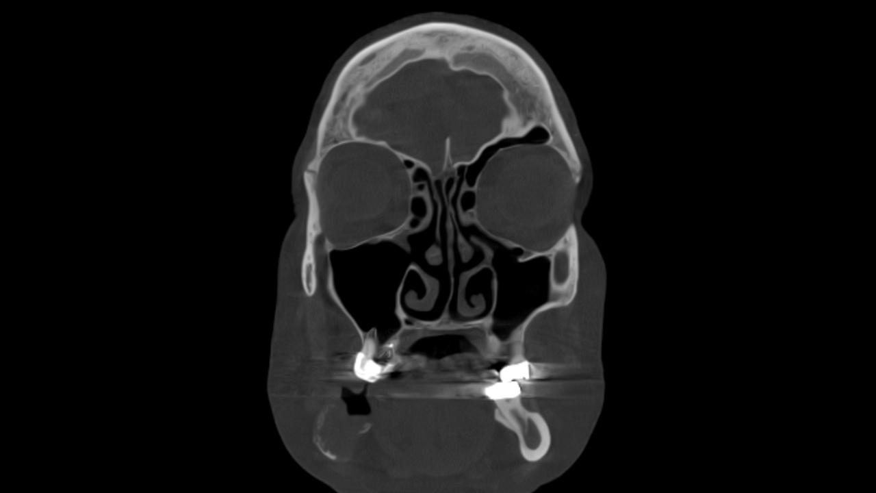
Study 3
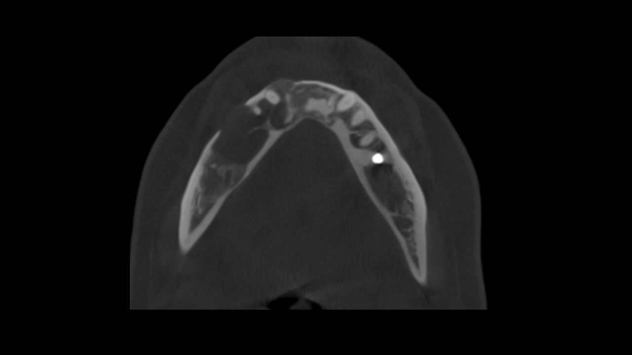
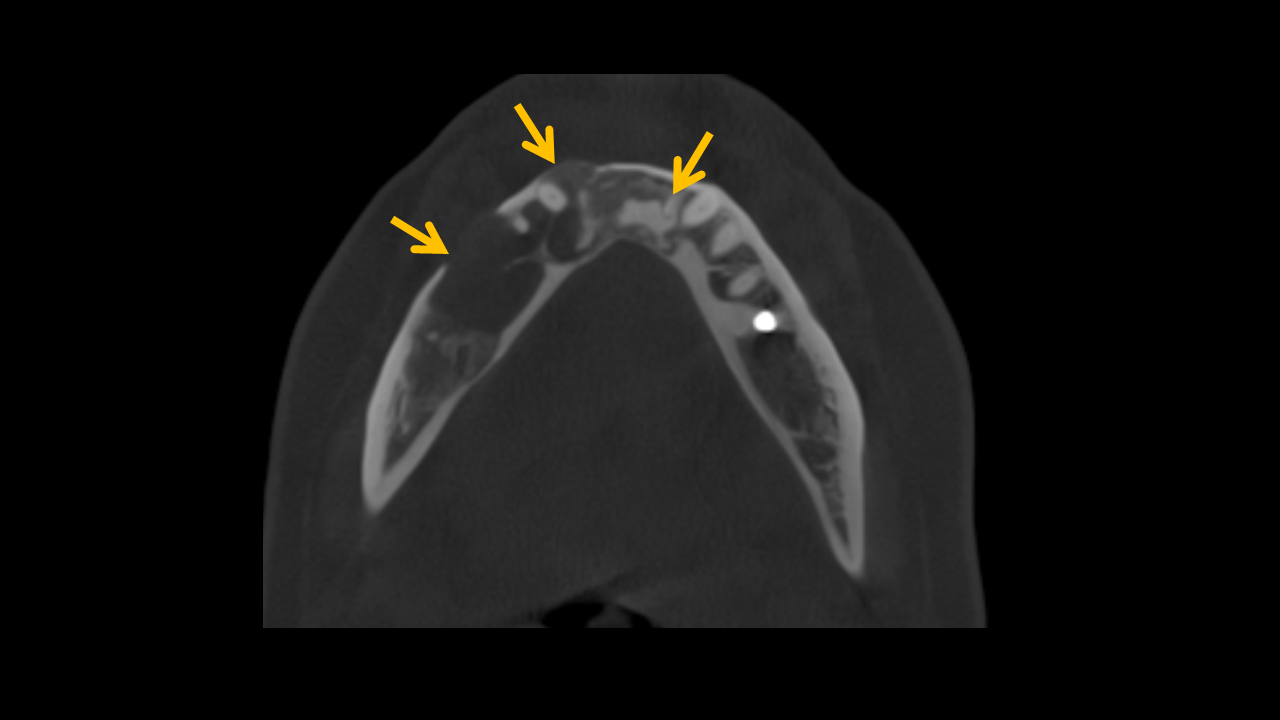
Finding: There is an osteolytic lesion involving the right mandibular arch, characterized by effacement and expansion of the buccal and lingual cortices. Notably, there is destruction of the alveolar crest, and the cortical boundaries of the mandibular canal are eroded. These imaging findings are highly suggestive of a malignant lesion.
Study 4
Finding: A mixed-density lesion is observed at the mandibular arch, characterized by varying levels of internal radiodensity surrounded by regions of low-density. The lesion has caused expansion and resorption of the adjacent cortical plates. No root resorption is detected in the associated teeth. These imaging findings are indicative of florid cemento-osseous dysplasia.
Let’s Connect!
We’re here to support your diagnostic imaging needs with precision, care, and efficiency.
Whether you're a referring provider, a patient, or simply have a question, we’d love to hear from you.
Please complete the form below and a member of our team will be in touch shortly.
This contact form is for general inquiries only. Please do not include any personal health information (PHI) in your message.